
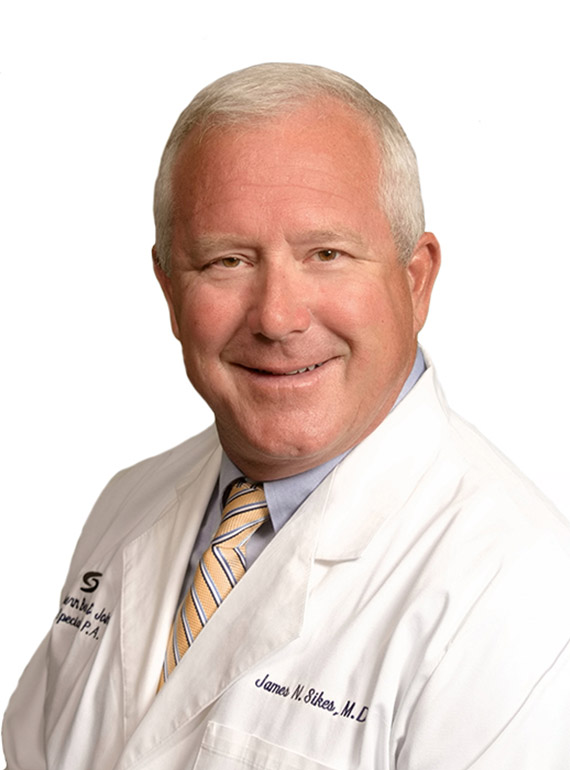
James Sikes was born the youngest of 4 children in Saraland, Alabama. His mother worked as a medical technologist and his father, who was a World War II veteran, was a dentist. After attending high school in Mobile, AL, he attended the University of Mississippi on an athletic scholarship. Following his undergraduate education he attended medical school at the University of Alabama Birmingham.
During medical school at UAB he performed research in total joint replacement at the Andrew’s South Highland Hospital and worked with local orthopedic surgeons. This experience led him to the orthopedic surgery training program in Jackson, MS, led by Dr. James Hughes and Dr. Bob McGuire. During his residency he chose a sabbatical in Jackson, Wyoming for further training in trauma and sports medicine.
After practicing for four years in Brookhaven, he joined Southern Bone & Joint Specialists in 2002. During Dr. Sikes’ years of service the practice has expanded to include the development of the Orthopedic Institute.
One of the unique offerings of Dr. Sikes’ practice is Bilateral Knee Replacement.
Direct Phone Number
(601) 543-7257
Robotic Assisted Surgery / Makoplasty. Over the years, joint replacement techniques and instrumentation have undergone countless improvements. Mako Technology is designed to help surgeons provide patients with a personalized surgical experience based on their specific diagnosis and anatomy.
- Mako Robotic-Arm Assisted Technology for Total Hip Replacement
- Mako Robotic-Arm Assisted Technology for Partial Knee Replacement
- Mako Robotic-Arm Assisted Technology for Total Knee Replacement
Services
Arthritis
The term arthritis literally means inflammation of a joint, but is generally used to describe any condition in which there is damage to the cartilage. Inflammation is the body’s natural response to injury. The warning signs that inflammation presents are redness, swelling, heat and pain.
The cartilage is a padding that absorbs stress. The proportion of cartilage damage and synovial inflammation varies with the type and stage of arthritis. Usually the pain early on is due to inflammation. In the later stages, when the cartilage is worn away, most of the pain comes from the mechanical friction of raw bones rubbing on each other.
There are over 100 different types of rheumatic diseases. The most common are:
Osteoarthritis: Also called degenerative joint disease, this is the most common type of arthritis and occurs often in older people. It affects cartilage, the tissue that cushions and protects the ends of bones in a joint. With osteoarthritis the cartilage starts to wear away over time. In extreme cases the cartilage can completely wear away, leaving nothing to protect the bones in a joint and causing bone-on-bone contact. Bones may also bulge, or form a bone spur at the end of a joint. Osteoarthritis causes joint pain and can limit a person’s normal range of motion. When severe, the joint may lose all movement, causing disability — most often when the disease affects the spine, knees and hips.
Rheumatoid Arthritis: An auto-immune disease in which the body’s immune system attacks healthy joints, tissues and organs. Occurring most often in women of childbearing age (15–44), it inflames the lining (synovium) of joints, causing pain, stiffness, swelling and loss of function. When severe it can deform a joint. It affects mostly the joints of the hands and feet and tends to be symmetrical — the same joints on both sides of the body at the same time. No other form of arthritis is symmetrical. About two to three times as many women as men have this disease.
Post-traumatic arthritis: Arthritis developing following an injury to the hand, wrist or elbow. The condition may develop years after trauma such as a fracture, severe sprain or ligament tear.
Psoriatic arthritis: Occurs in some people with psoriasis, a scaling skin disorder, affecting the joints at the ends of the fingers and toes. It can also cause changes in the fingernails and toenails. Back pain may occur if the spine is involved.
Causes of Arthritis
Osteoarthritis is caused by the wearing out of the cartilage covering the bone ends in a joint. This may be due to excessive strain over prolonged periods, or to other joint diseases, injury or deformity. Primary osteoarthritis is commonly associated with ageing and general degeneration of joints. Secondary osteoarthritis is generally the consequence of another disease or condition, such as repeated trauma or surgery to the affected joint, or abnormal joint structures from birth.
Rheumatoid arthritis is often caused when the genes responsible for the disease are triggered by infection or environmental factors. With this trigger the body produces antibodies against the joint, which may cause rheumatoid arthritis. Fractures at joint surfaces and joint dislocations may predispose an individual to post-traumatic arthritis. Uric acid crystal build-up is the cause of gout, and long-term crystal build-up in the joints may cause deformity.
Symptoms of Arthritis
Symptoms vary according to the form of arthritis, and each form affects the body differently. Arthritic symptoms generally include swelling and pain or tenderness in one or more joints for more than two weeks, redness or heat in a joint, limitation of motion, early morning stiffness, and skin changes including rashes.
Diagnosis
Doctors diagnose arthritis with a medical history, physical exam and X-rays of the affected part. Computed tomography (CT) scans and magnetic resonance imaging (MRI) scans are also performed to diagnose arthritis.
Treatment Options
There is no cure for arthritis. Your doctor may prescribe anti-inflammatory medicine and may recommend occupational therapy or physiotherapy, which includes exercises and heat treatment. In severe cases surgery may be suggested; the type will depend on your age and the severity of the disease. In the elderly with severe arthritis, joint replacement can give good results.
Initial treatment is conservative — rest, avoidance of vigorous weight-bearing activities, and non-narcotic analgesic and/or anti-inflammatory medications. With worsening symptoms a cane or braces may help. For more severe symptoms, a cortisone injection into the joint is frequently advised. When conservative measures have been exhausted and the condition has become disabling, surgery may be recommended. Options may include:
Synovectomy: Usually indicated for early inflammatory arthritis with significant swelling (synovitis) causing pain or limiting range of motion. It is the surgical removal of the inflamed synovium and may be performed using arthroscopy.
Arthroplasty: The surgeon removes the affected joint and replaces it with an artificial implant, usually when the joint is severely damaged by osteoarthritis, rheumatoid arthritis, post-traumatic arthritis or avascular necrosis. The goal is to relieve pain and restore normal function. It can be performed through an open or minimally invasive approach.
Arthrodesis: A fusion involving removal of the joint and fusing the bones together using metal wires or screws. Usually indicated when joints are severely damaged, when there is limited mobility, damage to surrounding ligaments and tendons, failed previous arthroplasty, or when heavy manual use is expected.
Your surgeon will discuss the options and help you decide which type of surgery is most appropriate for you.
Knee
Normal Anatomy of the Knee Joint
The knee is made up of four bones. The femur (thighbone) connects the hip to the knee. The tibia (shinbone) connects the knee to the ankle. The patella (kneecap) is the small bone in front of the knee that rides on the joint as the knee bends. The fibula is a shorter, thinner bone running parallel to the tibia on its outside. The joint acts like a hinge but with some rotation.
The knee is a synovial joint, lined by synovium, which produces fluid that lubricates and nourishes the inside of the joint. Articular cartilage is the smooth surface at the end of the femur and tibia; damage to this surface is what causes arthritis.
Femur — the largest and strongest bone in the body and the weight-bearing bone of the thigh. It provides attachment to most of the muscles of the knee.
Condyle — the two femoral condyles make up the rounded end of the femur. Their smooth articular surface allows the femur to move easily over the tibial meniscus.
Tibia — the second largest bone in the body and the weight-bearing bone of the leg. The menisci incompletely cover its superior surface where it articulates with the femur, acting as shock absorbers and assisting in rotation of the knee.
Fibula — although not weight-bearing, it provides attachment sites for the lateral collateral ligament (LCL) and the biceps femoris tendon. Its articulation with the tibia allows a slight degree of movement and flexibility.
Patella — attached to the quadriceps tendon above and the patellar ligament below, it rests against the anterior articular surface of the lower femur and protects the knee joint. It acts as a fulcrum for the quadriceps.
Menisci — the medial and lateral meniscus are thin C-shaped layers of fibrocartilage. Most of the meniscus has no blood supply, so when damaged it cannot undergo the normal healing process. The menisci act as shock absorbers and secondary stabilizers, interacting with the ligaments and most effective when those ligaments are intact.
Anterior Cruciate Ligament (ACL) — the major stabilizing ligament of the knee, located in the center and running from femur to tibia. It prevents the femur from sliding backward on the tibia and, together with the PCL, stabilizes the knee rotationally. Significant damage makes the knee unstable when planting the foot and pivoting.
Posterior Cruciate Ligament (PCL) — prevents the femur from moving too far forward over the tibia. It is the knee’s basic stabilizer, almost twice as strong as the ACL, and provides a central axis about which the knee rotates.
Collateral Ligaments — prevent hyperextension, adduction and abduction:
- Superficial MCL connects the medial epicondyle of the femur to the medial condyle of the tibia and resists valgus force.
- Deep MCL connects the medial epicondyle of the femur with the medial meniscus.
- LCL, entirely separate from the articular capsule, connects the lateral epicondyle of the femur to the head of the fibula and resists varus force.
Knee Fracture
A fracture is a break in the continuity of the bone. In younger individuals these fractures are caused by high-energy injuries such as a motor vehicle accident. In older people the most common cause is weak and fragile bone. Fractures of the knee can include:
Distal femur fracture: a break in the thighbone just above the knee joint, where the femur flares out like the mouth of a funnel.
Femoral shaft fracture: a break anywhere along the long, straight part of the femur.
Fractures of the proximal tibia: a break in the upper shin bone. Fractures that enter the knee joint may cause joint imperfections, irregular surfaces and improper leg alignment, which can lead to instability, arthritis and loss of motion. They can also injure surrounding soft tissues including skin, muscle, nerves, blood vessels and ligaments.
Tibial shaft fractures: a break along the length of the tibia between the knee and ankle joints, which can occur while playing sports such as soccer.
Diagnosis is made through medical history, physical examination and imaging. X-rays reveal whether the bone is intact or broken and the type and location of the fracture; a CT scan may be recommended to assess severity. Treatment may be non-surgical (skeletal traction, casts and braces) or surgical (internal or external fixation).
Internal Fixation
Intramedullary nailing: a specially designed metal rod is placed into the marrow canal of the femur, passed to the fracture site and secured with screws at both ends.
Plates and screws fixation: the surgeon repositions the broken bone ends and uses special screws or metal plates on the outer surface of the bone to hold the fragments in place.
External Fixation
Metal pins or screws are inserted into the femur and tibia and attached to a device outside the skin to hold bone fragments in place, allowing alignment and healing. If the bone is fractured in many pieces, a plate or rod maintains overall shape and length while it heals. In elderly patients where healing is delayed, a bone graft may be used. In severe cases the fragments are removed and the bone is replaced with a knee replacement implant.
The most common complications of surgery include infection, knee stiffness, delayed bone healing and knee arthritis.
Meniscus Tear
Meniscus tear is the most common knee injury in athletes, especially those in contact sports. A sudden bend or twist in the knee can tear the meniscus — a traumatic tear. Elderly people are more prone to degenerative meniscal tears as the cartilage wears out and weakens with age. The two wedge-shaped cartilage pieces between the thighbone and shinbone stabilize the knee joint and act as shock absorbers.
A torn meniscus causes pain, swelling, stiffness, and a catching or locking sensation that prevents full range of motion. Treatment depends on the type, size and location of the tear as well as your age and activity level. Small tears on the outer edge may be treated non-surgically; if symptoms persist, surgery may be recommended.
Surgical Treatment
Knee arthroscopy is the commonly recommended procedure. Options include meniscus removal (meniscectomy), meniscus repair and meniscus replacement. A tiny camera is inserted through a small incision so the surgeon can view inside the knee, and surgery is performed through other tiny incisions. During meniscectomy small shavers or scissors remove the torn tissue; in arthroscopic repair the torn meniscus is pinned or sutured. Meniscus replacement or transplantation replaces torn cartilage with tissue from a donor or a cultured patch, and is considered to relieve pain in patients who have undergone meniscectomy.
Mako Robotic-Arm Assisted Technology for Partial Knee Replacement
We understand that knowing what to expect from your joint replacement experience is important. As you read through this material, if you have additional questions please reach out to us.
Each patient is unique and can experience joint pain for different reasons. Pain from arthritis and joint degeneration can be constant or come and go, occur with movement or after rest, and be located in one spot or many. It is common to try medication and other conservative treatments first. If you haven’t experienced adequate relief, you may be a candidate for Mako knee replacement, which may provide relief from your knee pain.
How Mako Technology Works
Personalized Pre-operative Plan. Mako Robotic-Arm Assisted knee replacement is a treatment option for adults with mid to late-stage osteoarthritis of the knee. A CT scan of the diseased knee is uploaded into the Mako System software, where a 3D model is created and used to pre-plan and assist your surgeon.
Bone Preparation. In the operating room, your surgeon follows the personalized plan while preparing the bone for the Triathlon implant. Triathlon knee replacements are designed to work with the body to promote natural-like circular motion. The surgeon guides the robotic-arm to remove diseased bone and cartilage within the pre-defined area, and the system helps the surgeon stay within the planned boundaries.
The surgery is performed by an orthopedic surgeon who guides the robotic-arm to position the implant. The robotic-arm does not perform surgery, make decisions on its own, or move without the surgeon guiding it. The system also allows the surgeon to make adjustments during surgery as needed.
Important Information — Knee Replacements
General indications: Total knee replacement is intended for individuals with joint disease resulting from degenerative, rheumatoid and post-traumatic arthritis, and for moderate deformity of the knee.
Contraindications: Not appropriate for patients with certain infections, any mental or neuromuscular disorder that would create unacceptable risk of prosthesis instability or fixation failure, compromised bone stock, skeletal immaturity, or severe instability of the knee.
As with any surgery, knee replacement has serious risks including, but not limited to, nerve damage, circulatory compromise (including deep vein thrombosis), kidney failure, paralytic ileus, blood clots, blood loss or changes in blood pressure or heart rhythm, emboli, stroke, pneumonia, heart attack and death. Implant-related risks that may lead to revision include dislocation, loosening, fracture, nerve damage, abnormal bone formation, implant wear, metal sensitivity, soft-tissue imbalance, osteolysis and reaction to particle debris. Knee implants may not provide the same feel or performance as a normal healthy joint.
The information presented is for educational purposes only. Speak to your doctor to decide if joint replacement surgery is right for you. Individual results vary. The lifetime of a joint replacement is not infinite; strategies to prolong it include avoiding high-impact activities such as running and maintaining a healthy weight.
Stryker Corporation or its affiliates own or have applied for the trademarks Mako, Stryker and Triathlon. References on file (Mistry et al.; Piazza 2003; Wang et al. 2006; Gómez-Barrena et al. 2010; Hampp et al. 2017). MKOHMT-PE-3_Rev-1_13841. Copyright © 2017 Stryker Corporation.
Mako Robotic-Arm Assisted Technology for Total Knee Replacement
We understand that knowing what to expect from your joint replacement experience is important. As you read through this material, if you have additional questions please reach out to us.
Each patient is unique and can experience joint pain for different reasons. Pain from arthritis and joint degeneration can be constant or come and go, occur with movement or after rest, and be located in one spot or many. It is common to try medication and other conservative treatments first. If you haven’t experienced adequate relief, you may be a candidate for Mako Total Knee replacement, which may provide relief from your knee pain.
How Mako Technology Works
Personalized Pre-operative Plan. Mako Robotic-Arm Assisted Total Knee Replacement is a treatment option for adults with mid to late-stage osteoarthritis of the knee. A CT scan of the diseased knee is uploaded into the Mako System software, where a 3D model is created and used to pre-plan and assist your surgeon in performing the total knee replacement.
Bone Preparation. In the operating room, your surgeon follows the personalized plan while preparing the bone for the Triathlon Total Knee implant. Triathlon knee replacements are designed to work with the body to promote natural-like circular motion. The surgeon guides the robotic-arm to remove diseased bone and cartilage within the pre-defined area, and the system helps the surgeon stay within the planned boundaries.
The surgery is performed by an orthopedic surgeon who guides the robotic-arm to position the implant. The robotic-arm does not perform surgery, make decisions on its own, or move without the surgeon guiding it. The system also allows the surgeon to make adjustments during surgery as needed.
Important Information — Knee Replacements
General indications: Total knee replacement is intended for individuals with joint disease resulting from degenerative, rheumatoid and post-traumatic arthritis, and for moderate deformity of the knee.
Contraindications: Not appropriate for patients with certain infections, any mental or neuromuscular disorder that would create unacceptable risk of prosthesis instability or fixation failure, compromised bone stock, skeletal immaturity, or severe instability of the knee.
As with any surgery, knee replacement has serious risks including, but not limited to, nerve damage, circulatory compromise (including deep vein thrombosis), kidney failure, paralytic ileus, blood clots, blood loss or changes in blood pressure or heart rhythm, emboli, stroke, pneumonia, heart attack and death. Implant-related risks that may lead to revision include dislocation, loosening, fracture, nerve damage, abnormal bone formation, implant wear, metal sensitivity, soft-tissue imbalance, osteolysis and reaction to particle debris. Knee implants may not provide the same feel or performance as a normal healthy joint.
The information presented is for educational purposes only. Speak to your doctor to decide if joint replacement surgery is right for you. Individual results vary. The lifetime of a joint replacement is not infinite; strategies to prolong it include avoiding high-impact activities such as running and maintaining a healthy weight.
Stryker Corporation or its affiliates own or have applied for the trademarks Mako, Stryker and Triathlon. References on file (Mistry et al.; Piazza 2003; Wang et al. 2006; Gómez-Barrena et al. 2010; Hampp et al. 2017). MKOHMT-PE-3_Rev-1_13841. Copyright © 2017 Stryker Corporation.
Patellofemoral Instability
The knee can be divided into three compartments: patellofemoral, medial and lateral. The patellofemoral compartment is in the front of the knee between the kneecap and thigh bone. Patellofemoral instability means the patella (kneecap) moves out of its normal pattern of alignment, which can damage the underlying muscles and ligaments that hold the knee in place.
Causes
Instability can be caused by variations in the shape of the patella or its trochlear groove. Normally the patella moves up and down within the trochlear groove as the knee bends and straightens; instability occurs when it moves either partially (subluxation) or completely (dislocation) out of the groove. Contributing factors include:
Anatomical defect — flat feet, fallen arches or congenital abnormalities in the shape of the patella.
Abnormal Q angle — a high angle between the hips and knees, as in patients with knock knees.
Patellofemoral arthritis — loss of articular cartilage on the back of the kneecap.
Improper muscle balance — weak quadriceps can lead to abnormal tracking. Young, active individuals in sports are more prone to instability.
Symptoms
Pain when standing up from sitting and a feeling that the knee may buckle or give way. A partial or complete slip may cause severe pain, swelling, bruising, visible deformity and loss of function, and sometimes numbness or partial paralysis below the dislocation from pressure on nerves and blood vessels.
Diagnosis
Based on medical history and physical examination. X-rays, MRI and CT scans may be used to determine the cause and rule out other conditions.
Conservative Treatment
For partial dislocation (subluxation): pain medications, rest, ice, physical therapy, knee bracing and orthotics. A completely dislocated kneecap may need to be repositioned into the groove — a process called closed reduction.
Surgical Treatment
Surgery may be needed when non-surgical treatments fail, to realign the kneecap and decrease the Q angle. Procedures are broadly proximal (structures limiting outward movement are lengthened, or inner ligaments shortened) or distal (the Q angle is decreased by moving the tibial tubercle toward the inner knee). Under spinal or general anesthesia, an arthroscope guides evaluation and repair; a lateral retinacular release may be performed to allow the patella to sit properly in the femoral groove, and the medial tendons may be tightened. In severe malalignment, a tibial tubercle transfer (TTT) shifts and reattaches a section of bone using screws. Incisions are then closed with sutures.
Postoperative Care
Pain medications, leg elevation and ice packs reduce swelling. Crutches are used for the first few weeks and a knee immobilizer may stabilize the knee. A rehabilitation program supports recovery.
Risks and complications
- Loss of ability to extend the knee
- Recurrent dislocations or subluxations
- Arthrofibrosis (thick fibrous material around the joint)
- Persistent pain
Your surgeon will decide which procedure is appropriate for your particular situation.
Knee Osteoarthritis
Osteoarthritis, also called degenerative joint disease, is the most common form of arthritis and occurs most often in older people. It affects the cartilage covering the ends of bones in a joint; as the cartilage becomes damaged and worn, it causes pain, swelling, stiffness and restricted movement. It most commonly affects the hips, knees, hands and spine, and rarely the shoulders, wrists and feet.
Causes and Risk Factors
Caused by the wearing out of cartilage covering the bone ends, which may be due to being overweight, excessive strain over prolonged periods, previous fracture, growth abnormalities, joint diseases, injury or deformity. Some people have congenital joint abnormalities that cause early degeneration.
Diagnosis
Diagnosed with medical history, physical exam and X-rays. Your doctor examines the joint for swelling, pain, tenderness and range of motion; an X-ray may show loss of joint space and bone spur formation. There is no blood test for osteoarthritis.
Treatment
There is no known cure, but treatments and lifestyle changes can ease pain and symptoms, with the goal of reducing pain, improving movement and preventing further damage:
- Medications such as anti-inflammatory drugs, steroid injections and artificial joint fluid injections.
Lifestyle Modifications
- A moderate exercise program
- Heat or cold treatments
- A healthy, well-balanced diet
- Adequate rest
- Weight loss
- Protecting joints with assistive devices such as splints or braces
- Physical therapy — exercises to keep joints flexible and improve muscle strength
- Surgery — considered if nonsurgical treatment fails; joint replacement is an option when pain severely affects daily activities
Total Knee Replacement (TKR)
Total knee replacement, also called total knee arthroplasty, is a surgical procedure in which the worn or damaged surfaces of the knee joint are removed and replaced with artificial parts. Arthritis, injury or other joint diseases can damage the protective cartilage, causing extreme pain and difficulty with daily activities. Your doctor may recommend surgery if non-surgical options have failed.
Indications
Commonly indicated for severe osteoarthritis of the knee, in which joint cartilage gradually wears away. Your doctor may advise total knee replacement if you have:
- Severe knee pain that limits daily activities such as walking, rising from a chair or climbing stairs
- Moderate to severe pain during rest or that wakes you at night
- Chronic inflammation and swelling not relieved with rest or medications
- Failure to obtain relief from medications, injections, physical therapy or other conservative treatments
- A bow-legged knee deformity
Causes
The exact cause of osteoarthritis is not known, but commonly associated factors include injury or trauma to the joint, fractures at the knee, increased body weight, repetitive overuse, joint infection, inflammation and connective tissue disorders.
Diagnosis
Based on medical history, physical examination and X-rays, which typically show narrowing of the joint space in the arthritic knee.
Procedure
The goal is to relieve pain and restore alignment and function. Under spinal or general anesthesia, the surgeon exposes the joint, cuts the damaged femur at appropriate angles and attaches the femoral component. The damaged tibia and cartilage are shaved smooth and the tibial component is secured with bone cement or screws. A plastic articular surface is placed between the implants to provide smooth gliding movement and support body weight. The rear surface of the patella is prepared to receive a plastic component so it glides smoothly. The new joint is tested through its range of motion, irrigated and cleaned, and the incision is closed with drains and a sterile dressing.
Post-operative Care
Rehabilitation begins immediately. A physical therapist teaches exercises to strengthen the leg and restore movement. Knee immobilizers stabilize the knee, and you will walk with crutches or a walker. A continuous passive motion (CPM) machine may move the joint through a controlled range while you relax. A home exercise program strengthens thigh and calf muscles.
Risks and Complications
- Knee stiffness
- Infection
- Blood clots (deep vein thrombosis)
- Nerve and blood vessel damage
- Ligament injuries
- Patella (kneecap) dislocation
- Plastic liner wear
- Loosening of the implant
Total knee replacement is a safe and effective procedure to relieve pain, correct deformity and help you resume normal daily activities.
Bilateral Replacement
For patients with two painful arthritic knees, replacing both at the same time may be the best option. Under one anesthetic and one course of physical therapy, both knees are rehabilitated together. It is not twice the pain, but half the time to full recovery.
Over the last 15 years Dr. Sikes and Dr. Stonnington have refined the process by working together to lead two surgical teams in an orchestrated process of precision surgery, completing both total knee replacements rapidly. Skilled therapists coach this specially selected patient group to begin walking with a walker within hours after surgery.
The refined pathways of care — careful pre-op medical review, two-surgeon cooperation, rapid surgical time and multimodal pain management — have made this a particularly satisfying success story, with documented quality outcomes. To consider having both knees replaced may be an opportunity at a total victory rather than solving half the problem and being forced to complete the process again later.
Ligament Reconstruction (ACL)
The anterior cruciate ligament (ACL) is one of the major stabilizing ligaments of the knee — a strong, rope-like structure in the center running from the femur to the tibia. When it tears it does not heal, and often leads to a feeling of instability. ACL reconstruction is a commonly performed procedure that, with modern arthroscopic techniques, can be done with minimal incision and low complication rates.
ACL Reconstruction — Hamstring Tendon
This surgical procedure replaces the injured ACL with a hamstring tendon. The ACL connects the femur to the tibia, helps stabilize the knee, prevents excessive forward movement of the tibia relative to the femur, and limits rotational movements. A tear can make your knee feel unable to hold you up. Reconstruction rebuilds the torn ligament with a tissue graft.
Causes
An ACL injury most commonly occurs during sports involving twisting or overextending the knee:
- Sudden directional change
- Slowing down while running
- Landing from a jump incorrectly
- Direct blow to the side of the knee, such as during a football tackle
Symptoms
You might hear a loud “pop” and feel the knee buckle. Within a few hours the knee may swell from bleeding within the torn ligament. The knee may feel unstable or give way, especially when changing direction.
Diagnosis
Diagnosed with a thorough physical examination and tests such as X-rays, MRI scans and arthroscopy. The Lachman’s test may show increased forward movement of the tibia with a soft endpoint; the pivot shift test assesses the tear as the knee bends past 30°.
Procedure
The goal is to tighten the knee and restore stability. Under general anesthesia, two small cuts are made and an arthroscope is inserted; sterile solution expands the joint. The hamstring tendons are located, and two tendons are stripped from the muscle to prepare the graft. The torn ACL is removed, tunnels are drilled into the upper and lower leg bones, the graft is pulled through, and it is fixed with screws while it heals into the bone. Incisions are closed with sutures and a dressing applied.
Risks and Complications
- Numbness
- Infection
- Blood clots (deep vein thrombosis)
- Nerve and blood vessel damage
- Failure or loosening of the graft
- Decreased range of motion
- Crepitus (crackling or grating of the kneecap)
- Pain in the knee
- Repeat injury to the graft
Post-Operative Care
Rehabilitation begins immediately. A physical therapist teaches exercises to strengthen the leg and restore movement. Avoid competitive sports for 5 to 6 months to allow the graft to incorporate into the knee joint. ACL reconstruction is a very common and successful procedure, usually indicated for patients wishing to return to an active lifestyle.
ACL Reconstruction — Patellar Tendon
This procedure replaces the injured ACL with part of the patellar tendon, which connects the bottom of the kneecap to the top of the shinbone. Function and indications mirror the hamstring method — the difference is the graft source.
Procedure
Under general anesthesia, two small cuts are made and an arthroscope inspects the joint. The torn ACL is removed. The surgeon takes the middle third of the patellar tendon along with small bone plugs at each end; the remaining tendon is sutured back. Tunnels are drilled into the upper and lower leg bones, the graft is pulled through, and it is fixed with screws while it heals. Incisions are closed with sutures and a dressing applied.
Risks and Complications
Similar to the hamstring method: numbness, infection, blood clots, nerve and blood vessel damage, graft failure or loosening, decreased range of motion, crepitus, knee pain and repeat injury to the graft.
Post-operative Care
Rehabilitation begins immediately; avoid competitive sports for 5 to 6 months to allow the graft to incorporate. If you have injured your ACL, surgery may be needed to regain full function of your knee.
Hip
Normal Anatomy of the Hip Joint
The thigh bone (femur) and the pelvis (acetabulum) join to form the hip joint, a “ball and socket” joint. The “ball” is the head of the femur and the “socket” is the cup-shaped acetabulum. A smooth articular surface allows pain-free movement, and cartilage cushions the joint so the bones move smoothly. Because cartilage does not show on X-ray, a “joint space” appears between the femoral head and the acetabular socket.
Pelvis
A large, flattened, irregularly shaped bone, constricted in the center and expanded above and below. It consists of the ilium, ischium and pubis. The socket (acetabulum) is on the outer surface and joins the head of the femur to form the hip joint.
Femur
The longest bone in the skeleton. It joins the pelvis at the acetabulum to form the hip joint.
Femoro Acetabular Impingement (FAI)
FAI is a condition where excess friction in the hip joint from bony irregularities causes pain and decreased range of motion. The femoral head and acetabulum rub against each other, damaging the articular cartilage (the smooth surface of the ball or socket) or the labral tissue (the lining of the edge of the socket). Repeated friction frays or tears this tissue until, eventually, bone impacts on bone — commonly referred to as osteoarthritis. FAI generally occurs in two forms:
CAM impingement: the femoral head and neck are not perfectly round, most commonly due to excess bone, causing abnormal contact between surfaces.
PINCER impingement: the socket rim has overgrown and is too deep, covering too much of the femoral head and pinching the labral cartilage. It may also occur when the socket is abnormally angled backward. Most diagnoses include a combination of both forms.
Symptoms
- Groin pain associated with hip activity
- Pain in the front, side or back of the hip
- Dull ache or sharp pain
- Locking, clicking or catching sensation in the hip
- Pain in the inner hip or groin after prolonged sitting or walking
- Difficulty walking uphill
- Restricted hip movement
- Low back pain
- Pain in the buttocks or outer thigh
Diagnosis
Hip conditions should be evaluated by an orthopedic hip surgeon for proper diagnosis and treatment, including medical history, physical examination, and studies such as X-rays, MRI and CT scans.
Treatment Options
Conservative (non-surgical) management may not change the underlying abnormal biomechanics but can offer pain relief and improved mobility:
- Rest
- Activity modification and limitations
- Anti-inflammatory medications
- Physical therapy
- Injection of steroid and analgesic into the hip joint
Surgical Treatment
Hip arthroscopy to repair FAI is indicated when conservative measures fail to provide relief.
Hip Fracture
A hip fracture is a break near the hip in the upper part of the femur. The upper femur has two bony processes — the greater and lesser trochanters. Fractures can occur in the femoral neck, in the area between the greater and lesser trochanters, or below the lesser trochanter.
Hip fracture is most frequently caused by minor trauma in elderly patients with weak bones, and by high-energy trauma in young people. Long-term use of certain medicines, such as bisphosphonates for osteoporosis, increases the risk.
Signs and Symptoms
- Pain in the groin or outer upper thigh
- Swelling and tenderness
- Discomfort while rotating the hip
- Shortening of the injured leg
- Outward or inward turning of the foot and knee of the injured leg
An X-ray is used to diagnose a hip fracture; an MRI may also detect it. Depending on the area involved, hip fractures are classified as intracapsular, intertrochanteric or subtrochanteric.
Fractures may be corrected non-operatively or operatively. Traction may be an option if you are not fit for surgery: under local anesthesia, screws, pins and wires are inserted into the femur and a pulley system bears heavy weights to correct misaligned bones until the injury heals. Surgically, hip fractures can be treated with external fixation, intramedullary fixation, or plates and screws.
Mako Robotic-Arm Assisted Technology for Total Hip Replacement
We understand that knowing what to expect from the joint replacement experience is important. As you read through this material, if you have additional questions please reach out to us.
Each patient is unique and can experience joint pain for different reasons. If conservative treatments haven’t provided adequate relief, you may be a candidate for Mako Total Hip replacement, which may provide relief from your hip pain.
Mako Robotic-Arm Assisted Technology provides a personalized surgical plan based on your unique anatomy. A CT scan of the diseased joint is uploaded into the Mako System software, where a 3D model of your hip is created and used to pre-plan and assist your surgeon. In the operating room, the surgeon follows the plan while preparing the bone for the implant, guiding the robotic-arm within the pre-defined area. The system helps the surgeon stay within the planned boundaries for more accurate placement and alignment of the implant.
Mako Total Hip replacement is intended for patients who suffer from non-inflammatory or inflammatory degenerative joint disease, including osteoarthritis, post-traumatic arthritis, rheumatoid arthritis, avascular necrosis and hip dysplasia. The procedure follows a personalized plan built from the hip CT scan.
The surgery is performed by an orthopaedic surgeon who guides the surgeon-controlled robotic-arm to position the implant. The robotic-arm does not perform surgery, make decisions on its own, or move without the surgeon guiding it. The system also allows the surgeon to make adjustments during surgery as needed.
Important Information — Hip Replacements
Hip joint replacement is intended for individuals with joint disease from degenerative and rheumatoid arthritis, avascular necrosis, fracture of the neck of the femur, or functional deformity of the hip. It is not appropriate for patients with certain infections, mental or neuromuscular disorders creating unacceptable risk, compromised bone stock, skeletal immaturity, severe instability, or excessive body weight.
Like any surgery, joint replacement has serious risks including, but not limited to, pain, bone fracture, change in treated leg length, joint stiffness, joint fusion, amputation, nerve damage, circulatory compromise (including deep vein thrombosis), kidney failure, paralytic ileus, blood clots, blood loss or changes in blood pressure or heart rhythm, emboli, stroke, pneumonia, heart attack and death. Implant-related risks that may lead to revision include dislocation, loosening, fracture, nerve damage, abnormal bone formation, implant wear, metal sensitivity, soft-tissue imbalance, osteolysis, audible sounds during motion and reaction to particle debris.
The information presented is for educational purposes only. Speak to your doctor to decide if joint replacement surgery is appropriate for you. Individual results vary and the lifetime of any joint replacement is limited. Strategies to prolong it include avoiding high-impact activities such as running and maintaining a healthy weight.
Stryker Corporation or its affiliates own or have applied for the trademarks Mako and Stryker. References on file (Nawabi et al. 2013; Illgen 2014). MKOHMT-PE-3_Rev-1_13841. Copyright © 2017 Stryker Corporation.
Total Hip Replacement (THR)
Total hip replacement removes damaged cartilage and bone from the hip joint and replaces it with artificial components. The hip is a ball-and-socket joint between the femoral head (ball) and the pelvic acetabulum (socket), covered by smooth articular cartilage that cushions and enables smooth movement. Several conditions can damage this cartilage; total hip replacement is an option to relieve severe arthritis pain that limits daily activities.
Disease Overview
Arthritis causes pain, swelling, stiffness and limited movement. The three most common types affecting the hip are:
Osteoarthritis: progressive wearing away of joint cartilage; as it wears down, the bone ends rub together and cause pain.
Rheumatoid arthritis: an autoimmune disease in which the synovium becomes inflamed and produces excess joint fluid, leading to loss of cartilage, pain and stiffness.
Traumatic arthritis: results from a hip injury or fracture that damages the cartilage.
Symptoms
The most common symptom is joint pain and stiffness with limited range of motion. Vigorous activity can increase pain and stiffness and may cause limping.
Diagnosis
Made by evaluating medical history, physical examination and X-rays.
Surgical Procedure
Recommended when conservative options fail. Under general anesthesia, the surgeon exposes the hip and dislocates the femur from the acetabulum. The socket is cleaned and the damaged bone removed with a reamer; the acetabular component is inserted with screws or bone cement, and a liner of plastic, ceramic or metal is placed inside. The femur is prepared to fit the metal femoral component, which is inserted by press fit or bone cement. The femoral head component is placed on the stem, the parts are secured, and the muscles and tendons are repaired before closing the incision.
Post-operative Care
To prevent dislocation and ensure healing:
- Avoid the combined movement of bending your hip and turning your foot inward
- Keep a pillow between your legs while sleeping for 6 weeks
- Never cross your legs or bend your hips past a right angle (90°)
- Avoid sitting on low chairs
- Use a grabber instead of bending down to pick up things
- Use an elevated toilet seat
Risks
- Infection
- Dislocation
- Fracture of the femur or pelvis
- Injury to nerves or blood vessels
- Blood clots in the leg veins
- Leg length inequality
- Hip prosthesis wear
- Failure to relieve pain
- Scar formation
- Pressure sores
Total hip replacement is one of the most successful orthopedic procedures for patients with hip arthritis, relieving pain, restoring function and improving quality of life.
Computer Navigated Surgery
Computer navigation is a modern advance in orthopedic surgery used for replacement of arthritic joints. It provides the surgeon with real-time 3-D images of the mapped joint and the surgical instruments during surgery. Infrared sensors fixed to the bones and instruments are tracked by an infrared camera above the surgical table connected to the computer, which generates real-time images to guide the surgeon in precisely resurfacing and cutting the bone and fixing the implant according to the pre-operative plan.
The surgery is still performed by the surgeon — computer navigation is a tool that guides and improves the outcome, and cannot replace the skills of an experienced surgeon. Precise alignment of the implant can help extend the life of the implant.
Direct Anterior Hip Replacement
Anterior hip replacement is a minimally invasive, muscle-sparing alternative to traditional hip replacement. Traditionally the incision is made on the side (lateral) or the back (posterior) of the hip, both of which involve cutting major muscles. With the anterior approach the incision is made in front of the hip, letting the surgeon access the joint without cutting muscles. A special operating table facilitates the anatomical positions needed to replace the hip anteriorly.
Potential benefits compared with traditional hip replacement may include:
- Smaller incision
- Minimal soft tissue trauma
- Reduced post-op pain
- Less blood loss
- Shorter surgical time
- Faster healing time
- Less scarring
- Earlier mobilization
- Fewer post-operative restrictions
- Reduced hip dislocations
- Decreased hospital stay
After a traditional hip replacement, restrictive hip precautions are usually required (avoiding combined bending and inward turning of the foot, sleeping with a pillow between the legs for 6 weeks, not crossing the legs or bending past a right angle, avoiding low chairs, using a grabber and an elevated toilet seat). For anterior hip replacement patients, however, these precautions are generally unnecessary. Because the muscles are not cut, the risk of dislocation is greatly reduced, allowing more freedom of movement and faster rehabilitation.
Sports Medicine
Sports Medicine Overview
Sports injuries occur while playing indoor or outdoor sports or exercising. They can result from accidents, inadequate training, improper use of protective devices, or insufficient stretching or warm-up. The most common are sprains and strains, fractures and dislocations. The most common treatment for injury is rest, ice, compression and elevation (RICE):
- Rest: avoid activities that may cause injury.
- Ice: apply ice packs over a towel to the affected area for 15–20 minutes, four times a day for several days. Never place ice directly on the skin.
- Compression: use elastic wraps, air casts or splints to reduce swelling.
- Elevation: raise the injured part above heart level to reduce swelling and pain.
Measures to help prevent sports injuries include following a strengthening program, gradually increasing exercise level, wearing properly fitted protective gear, warming up and cooling down, avoiding exercise right after a large meal, maintaining a healthy diet, avoiding play when injured or tired, learning the rules of the game, and ensuring you are physically fit to play.
Hip Injuries
Femur fractures, labral tears and hip dislocation are common. The hip bears more weight and is susceptible to injury during sport, requiring prompt medical intervention. Rehabilitation and physical therapy are often recommended.
Knee Injuries
The ACL may tear with overuse; it heals poorly and may cause instability. Cartilage damage and meniscal tears are also common. Treatment may require open or minimally invasive surgery followed by physical therapy.
Foot and Ankle Injuries
Common in sports such as football, hockey and skating. Treatment may include orthotics, braces, physical therapy, injections or surgery. Common injuries include sprains and strains, ankle fractures and Achilles tendinitis.
Shoulder Injuries
Severe shoulder pain in sports such as tennis, basketball and gymnastics may result from a torn ligament or shoulder dislocation, often from overuse. Simple or acute injuries may be treated conservatively; chronic injuries may require surgery.
Shoulder Arthroscopy
Arthroscopy is a minimally invasive diagnostic and surgical procedure for joint problems. Shoulder arthroscopy uses a pencil-sized instrument called an arthroscope, with a light and camera that projects images to a screen. It treats conditions and injuries involving the bones, cartilage, tendons, ligaments and muscles of the shoulder.
Disease Overview
The shoulder is a ball-and-socket joint where the head of the humerus articulates with the glenoid of the scapula. Cartilage prevents friction between the bones; tendons and ligaments provide strength and stability. Injury or disease can make the joint unstable, causing pain, inflammation and reduced mobility.
Indications
Indicated when conservative treatment fails to relieve pain and disability, for conditions such as:
- Shoulder impingement
- Rotator cuff tear
- Frozen shoulder or stiffness
- Shoulder instability
- Biceps rupture
- Damaged cartilage or ligaments
- Bone spurs or bony projections
- Arthritis of the collarbone
Procedure
Performed under general or regional anesthesia. Sterile fluid expands the joint for a clear view; a button-sized hole admits the arthroscope, and surgical instruments enter through separate small holes to remove and repair damage. Incisions are closed with stitches or sterile bandage strips.
Post-operative Care
Small wounds heal within a few days. Pain medications keep you comfortable, the arm is placed in a sling for a short period, and physical therapy improves mobility and strength.
Advantages
- Less pain
- Fewer complications
- Shorter hospital stay
- Faster recovery
Risks and Complications
Include infection, bleeding, damage to nearby nerves or blood vessels, delayed healing and, in some cases, stiffness of the shoulder joint. Active participation in physical therapy helps prevent stiffness.
Rotator Cuff Repair
The rotator cuff is a group of tendons in the shoulder that provide support and enable a wide range of motion. Major injury may tear these tendons — a rotator cuff tear — one of the most common causes of shoulder pain in middle-aged and older adults. It may occur with repeated overhead activity, during sports or in motor accidents, causing severe pain, arm weakness, and a crackling sensation, along with possible stiffness, swelling, loss of movement and tenderness at the front of the shoulder.
Repair
Rotator cuff tears are best viewed on MRI. Symptomatic relief may come from conservative treatment — rest, a shoulder sling, pain medications, steroid injections and certain exercises — but surgery is required to reattach the tendon to the bone. Repair may be performed by open surgery or, more commonly today, arthroscopically through tiny incisions of about 1 cm each, using suture anchors that attach the tendon to the shoulder bone. Following surgery you may be advised to practice motion and strengthening exercises.
The benefits of arthroscopy compared with open shoulder surgery include:
- Smaller incisions
- Minimal soft tissue trauma
- Less pain
- Faster healing time
- Lower infection rate
- Less scarring
- Earlier mobilization
- Usually performed as outpatient day surgery
Joint Restoration
A joint connects two or more bones, providing support and enabling movement of the knee, hip, elbow or shoulder. A diseased or injured joint results in pain and interferes with movement. Osteoarthritis, rheumatoid arthritis, bursitis, strains, sprains and other injuries can lead to painful joints, stiffness and swelling. Joint restoration repairs injured joints, slows the progression of arthritis and restores the joint’s original function.
Joint restoration initially takes a conservative approach to relieve pain and promote healing — rest, modifying activities, physical therapy and medications. Surgery may be needed for those who do not benefit from non-surgical treatment, and may involve joint reconstruction or joint replacement.
Joint Reconstruction
Ranges from minor joint repairs to total joint replacement, chosen based on the severity of the disorder. Procedures include cartilage restoration, ligament and tendon repair, osteotomy, joint resurfacing and total joint replacement:
- Osteotomy: cutting of the bone, performed for early-stage osteoarthritis to shift weight off the damaged part of the joint, relieving pain and improving function.
- Tendon repair: suture anchors sew the tendons to the kneecap, restoring the original function of the joint and surrounding tissues.
- Resurfacing surgery: hip resurfacing replaces and caps the damaged hip ball with a metal prosthesis rather than fully replacing it — recommended for younger patients, with a lower chance of dislocation than total hip replacement.
Joint Replacement
Replaces a damaged joint with an artificial one, replacing only the damaged parts. The prosthesis can be plastic, metal or both, and cemented, non-cemented or a combination. A minimally invasive approach uses smaller, less-invasive incisions that avoid disrupting surrounding muscles and soft tissues, with advantages of less muscle dissection, minimal pain, quick recovery and faster rehabilitation.
Patient Information
Patient Forms
With the increasing complexity of medical information compliance, a medical assistant will be assigned to work with each patient to enter necessary medical facts and insurance information into your medical file. This process takes 15–25 minutes at your first visit. To expedite it, please bring a list of medications and be prepared to answer questions about your medical history. As a courtesy, the staff will file insurance claims with your medical insurance company, and the business office staff is available to answer questions about your policy or office charges.
Preparing for Surgery & Procedure
Preparing for Surgery
Once you and your doctor decide that surgery will help you, you will need to learn what to expect and create a treatment plan for the best results afterward. Preparing mentally and physically is an important step toward a successful result, and understanding your role will help you recover more quickly with fewer problems.
Working with Your Doctor
Before surgery your doctor performs a complete physical examination to rule out conditions that could interfere with surgery or outcomes. Routine tests such as blood work and X-rays are usually done about a week before major surgery. Recommendations include:
- Discuss all medications with your doctor and family physician to learn which to stop before surgery
- Discuss options for potential blood replacement, including donating your own blood
- If overweight, lose weight before surgery to decrease stress on your new joint — but do not diet during the month before surgery
- Stop aspirin, anti-inflammatory medications, warfarin or other drugs that increase bleeding one week before surgery
- If you smoke, stop or cut down to reduce risks and improve recovery
- Have any tooth, gum, bladder or bowel problems treated to reduce infection risk
- Eat a well-balanced diet, supplemented by a daily multivitamin with iron
- Report any infections — surgery cannot be performed until they have cleared
- Arrange help with everyday tasks like cooking, shopping and laundry
- Put frequently used items within easy reach to avoid reaching and bending
- Remove loose carpets and tape down electrical cords to avoid falls
- Have a stable chair with a firm seat cushion, firm back and two arms
Preparing for the Procedure (Day Surgery)
- Have someone available to drive you home — you will not be able to drive for at least 24 hours
- Do not drink or eat anything in the car on the trip home
- Anesthesia, food and car motion can cause nausea — wait until you are hungry, then start with a light meal and avoid greasy food for the first 24 hours
- If you had surgery on an extremity, keep it elevated and use ice as directed to decrease swelling and pain
- Take pain medicine as directed — begin as you start getting uncomfortable, before the pain is severe
Post-Op Instructions
Apart from the specific instructions for your particular surgery, general instructions to follow afterward are:
- Take pain-relieving and other medications as advised, with food. After the first 48 hours, take pain medication only when needed.
- Do not drink alcohol, drive, operate machinery or sign a legal document for the first 24 hours after surgery, as the effect of the sedative and/or anesthesia may last that long.
- Use ice packs to control swelling, making sure the ice bag does not leak into the dressing. Ice packs can be used liberally for 2 weeks and later if required.
- Follow the specific activity restrictions advised. It is easier to prevent pain than to manage it once developed. Rest for a few days and keep the operated extremity elevated above heart level to control swelling.
- You will be instructed when the surgical site may be washed briefly with an antibacterial soapy wash rag, then thoroughly dried and covered with dry gauze and no ointment.
- Dr. Sikes will instruct you when to begin physical therapy. He prefers that you use the therapy team at Southern Bone & Joint for your recovery.
- Eat a healthy diet and drink plenty of non-alcoholic, non-caffeinated drinks.
- Schedule your follow-up appointment as advised.
Please consult your doctor immediately if you experience any of the following:
- Increased drainage from the incision
- Increased redness around the operated area
- Increased swelling that does not decrease with ice and elevation
- Foul odor
- Fever greater than 101°F
- Coldness, numbness or blanched white or bluish color of the fingers or toes
- Sudden calf pain or shortness of breath
- Chest pain
Useful Links
Appointments, Portal & Billing
Schedule an appointment, access the secure patient portal, or pay your bill online: